February 26, 2025
Most people have a basic understanding of magnetic resonance imaging (MRI) — you lie still inside a tube while the surrounding machine uses a magnetic field and radio waves to create detailed images of your internal tissues. But not all MRI scanners are created equally.
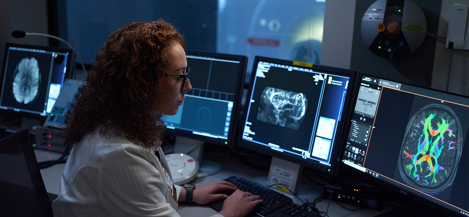
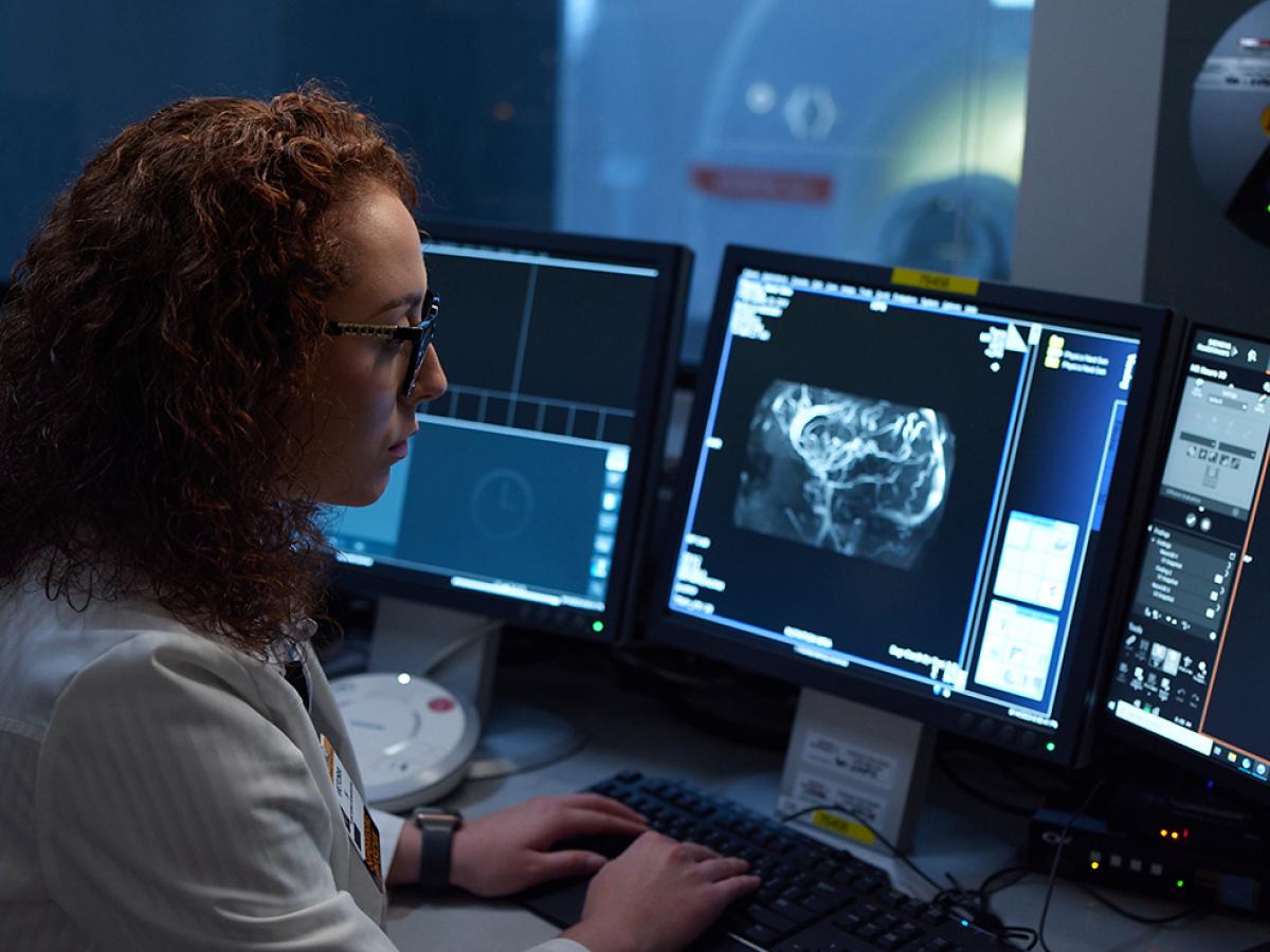
Currently, the most powerful MRI available to patients in North America is the 7-Tesla (7T) MRI, and it’s allowing MU Health Care researchers and providers to see inside the brain and other parts of the body like never before.

“When you get a better look at what’s going on inside, you start to understand the mechanisms of a disease,” says MU Health Care neuroradiologist Dr. Mai-Lan Ho. “It opens the door to interesting discussions and more treatment possibilities.”
Dr. Ho is vice chair of radiology and director of neuroradiology at MU Health Care. One of her primary responsibilities is making sure that neurologists, epileptologists (epilepsy specialists) and neurosurgeons understand the care opportunities they have with the current and future capabilities of the 7T MRI. Located in the NextGen Precision Health Building, the 25-ton Siemens Healthineers MAGNETOM Terra 7-Tesla MRI scanner is one of fewer than 20 such scanners in North America — and MU Health Care is charting the course for its use in clinical care.
What Is 7-Tesla MRI?
MRI field strength is measured in units of tesla (T). Higher tesla means a stronger magnetic field, which can create sharper and more detailed images of small anatomy.
To put it in perspective, standard MRI is performed on a 1.5T or 3T machine. 7T scanners are much stronger, enabling medical professionals to see brain abnormalities that weren’t previously visible. Some of our providers say it’s like comparing a smartphone camera from 10 years ago to the camera on a new phone.
“7T MRI is still just one piece of the puzzle when it comes to understanding neurological diseases like epilepsy,” Dr. Ho says. “But it helps us visualize lesions with more clarity and confidence and understand more about the underlying disease process.”
What 7T MRI Imaging Means for Patients
The Food and Drug Administration (FDA) cleared the Siemens Healthineers MAGNETOM Terra 7T MRI for brain and knee imaging in 2017. While most of the 7T MRI machines in the United States are being used for clinical and research imaging, with a few for research only, MU Health Care — which acquired a 7T MRI scanner in 2020 — focuses on using this powerful technology for patient benefit.
“We do a good amount of research and investigation using the machine,” Dr. Ho says. “But we are also pioneering the use of first-line clinical 7T imaging in our region. That means patients don’t need to do a regular MRI in order to be referred for 7T MRI.”
It’s not that other institutions don’t want to use the 7T. Insurance covers 7T MRI the same way as other MRI procedures. Rather, knowing how to use and adjust the machine can be quite difficult.
“It's the type of techniques and all the factors that go into making an image. You’ve got more energy, more sensitivities; it’s all about finding the right balance of time, physics and energy to make it work,” says James “Chris” Jones, a 7T MRI technician at MU Health Care. “We've been able to not only figure it out, but use it practically. We're making it work clinically, and we're seeing the differences.”
It’s important to note imaging with a 7T MRI can take slightly longer than regular MRI scanners, and not every patient is a candidate. Jones and his team have used 7T MRI for many conditions, including headaches and pituitary diseases. But he says 7T imaging truly shines and offers life-changing benefits when used for:
Epilepsy
As a Level IV epilepsy center, MU Health Care treats the most complex cases and offers the highest level of care, but there are still challenges. Standard imaging does not always reveal the underlying cause of seizures. 7T MRI scans are so detailed that they can better identify abnormal brain anatomy and help clinicians create a more effective treatment plan.
“Some patients have had seizures their whole lives,” Jones says, “and this technology is changing their outcomes.”
Metastatic Cancer
When cancer starts in or spreads (metastasizes) to the brain, an accurate picture of the diseased areas gives providers better understanding of the extent of disease and how to best treat it. Jones reports that brain tumors are among the top three most common uses for 7T MRI at MU Health Care.
“We had a patient scanned with standard MRI and the images showed three areas of metastatic cancer,” Jones says. “When we scanned her with 7T MRI, we saw nine. Those details make a big difference.”
Multiple Sclerosis
Misdiagnosis of multiple sclerosis (MS) is a common problem. During the diagnostic process, providers use imaging to view plaques (scars) inside the brain. But the best way to confirm those plaques indicate MS is by identifying the central vein sign (CVS) — a blood vessel that runs through the center of a plaque.
“We’ve never been able to image the CVS and see it clearly,” Jones says. “But with 7T MRI, we can see it. Now, when a patient comes in with plaques in their brain, we can make a more definitive diagnosis.”
Musculoskeletal Issues
MU Health Care is one of just 11 DNV designated Orthopedic and Spine Centers of Excellence in the nation. With the addition of 7T MRI scanning, we can find more joint abnormalities and understand how to treat them.
“We’ve had orthopedic surgeons look at our knee scans and notice things they couldn’t see before,” Jones says. “It changes their diagnosis and gives them the ability to be more precise with their plan.”
7T MRI machines currently include equipment for imaging knees. But our providers are thinking outside of the box to image other small joints.
“We’ve been able to image wrists, hands, fingers and elbows,” Jones says. “We’ve done it, following our organization’s procedures, including Institutional Review Board oversight, and we’re becoming more proficient at it in the hopes that 7T MRI will become front-line imaging for musculoskeletal conditions.”
How MU Health Care is Leading the Way for 7T Imaging
Bringing 7T MRI into clinical practice isn’t as simple as choosing to offer it to patients. The process involves research, partnerships, advanced expertise and a commitment to ensuring 7T MRI is safe and effective for patients.
We’ve taken several steps to offer 7T MRI to patients:
Achieving ACR Accreditation for 7T MRI
In the fall of 2024, MU Health Care achieved accreditation through the American College of Radiology (ACR) for 7T MRI. The ACR considers its accreditation process to be the “gold standard in medical imaging.” Currently, fewer than 10 7T MRI scanners in the United States have achieved it.
“We pursued accreditation early on because our 7T MRI scanner sees a higher level of clinical use than most 7T MRI machines in the United States,” says Bill Keller, MU Health Care manager of radiology services. “The accreditation ensures we are meeting and maintaining quality and safety standards for patient care.”
Leveraging Academic Research
The 7T system has some challenges. Compared to 1.5T and 3T scanners, it’s more sensitive to patient movement, metal implants, and technical issues. As a result, 7T images tend to show more artifacts — visual distortions to images. The 7T hardware is also complex and requires specific expertise to realize and harness its full potential.
As an academic health system, MU Health Care has worked with researchers at the University of Missouri to improve the 7T technology. A major turning point was finding a way to perform 7T imaging with our research parallel transmission (pTx). This cutting-edge imaging technique is difficult to master in high field MRI, but greatly improves image quality and decreases artifacts. MU Health Care has experts who have worked to optimize the 7T MRI settings for pTx imaging, and has scanned over 300 patients using this technique.
“Our academic team tested and optimized this imaging technique so that we can get the best image quality possible,” says Jones. “Thanks to them, we’re now using our research pTx in the clinical setting for neurology, and we’re seeing a difference in image quality and speed.”
Partnering for Success
In 2019, MU Health Care and the University of Missouri System entered a value partnership with Siemens Healthineers — a global pioneer in medical technology and the manufacturer of the MAGNETOM Terra 7T MRI scanner. The 10-year partnership — called the Alliance for Precision Health — offers our researchers and providers access to the newest technology and the scientists who create it.
“This collaboration provides an opportunity for our research scientists to access some of the newest equipment from Siemens Healthineers and understand their development processes,” Keller says. “On our end, we share our clinical experience with 7T MRI so they can incorporate our feedback into their research.” Through the partnership with Siemens Healthineers, we identify challenges and introduce solutions that in turn affect how other organizations are using 7T MRI worldwide.
Looking Ahead With 7-Tesla MRI
Research conducted with 7T MRI is ongoing, focusing on areas and diseases including:
- Brain tumors
- Epilepsy
- Multiple sclerosis
- Dementia
- Traumatic brain injury
- Vascular disease
- Headache
- Joint problems
We have only uncovered the tip of the iceberg for what can be done with 7T MRI. The technology and functionality will continue to improve with each new upgrade of the machine’s software and hardware.
“We continue to expand our range, both in research and clinical use,” Keller says. “This technology is promising, and we’re doing everything we can to bring it to patients.”
Next Steps and Useful Resources
- Want to learn more about imaging at MU Health Care? See the options.